Home » Ureteroscopy And Ureterorenoscopy Urs
A ureteroscopy (URS) is a simple investigation which allows your surgeon to make a diagnosis and give treatment where necessary. It involves using either a rigid telescope called a ureteroscope or a flexible one called a ureterorenoscope.
Urine is made bythe kidneys then drains into your bladder via narrow pipes called ureters where it is stored until you empty itby urinating through your urethra or “water pipe”.
A ureteroscopy allows the surgeon to look up into the ureter and/or the kidney. There are different reasons for having a ureteroscopy (see below). Your surgeon will explain why you need to have this and what to expect before, during and after the procedure.
A ureteroscope is a thin telescope (‘scope’). It is passed by a surgeon through the urethra (water pipe) into the bladder then up into the ureter. The doctor can look down the scope and can see images on a monitor. There are two types of ureteroscopes:
Both types of ureteroscope have side channels through whichvarious thin devices can pass. For instance the doctor may take a small tissue sample (biopsy) from the lining of the ureter or kidney with a thin ‘grabbing’ instrument, passed down a side channel. Additionally the doctor may use laser to fragment stones or vaporise small tumours and a variety of instruments to extract stones.
A ureteroscopy takes approximately 10 – 90 minutes to perform and can often be done as a ‘day case’, which means that you may be able to go home shortly afterwards.
Give common reasons why this treatment is recommended.
Ensure this section is realistic and does not give false hope.
Detail medical benefits and the improvements in mobility, social functioning, prognosis, etc. that can be expected
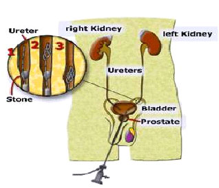
Diagram 1: A ureteroscope showing the basket stone removal in a male patient
Your doctor will pass the scope up the urethra (water pipe) and then up into the ureter to examine the inside of the ureter.
The doctor may need to take a biopsy (tissue sample). A small piece of tissue can be removed and sent to a laboratory for examination.
If there is any blockage or a stone in the ureter this can also be removed via the scope (see diagram) using “basket” or stone grasping instruments.
You will also need a ureteroscopy for insertion, removal or changing of a stent. This is a soft plastic tube placed between the kidney and the bladder. This tube will ensure urine can flow freely down the ureter into the bladder via the stent. The procedure is usually performed under X-ray control. Stents may be temporary or long term. Some temporary stents may have a string attached to the end to allow them to be removed while those that do not have the string will have to be removed or changed under anaesthetic.
To help with diagnosis
A ureteroscopy can help find the cause of symptoms such as:
To treat certain conditions or to do certain procedures
By using various instruments passed down the side channels of the scope your doctor can:
All treatments and procedures have risks and we will talk to you about the risks of having a ureteroscopy when you are seen in clinic.
The complications or side effects which can arise include:
Common (greater than 1 in 10)
Occasional (between 1 in 10 and 1 in 50)
Rare (less than 1 in 50)
If you require a general anaesthetic there are a number of issues that affect the chances of suffering complications, including: age, weight, lifestyle issues and your general state of health. Your anaesthetist and/or your surgeon can give further details. The information below on risks is provided by the Royal College of Anaesthetists.
Very common (1 in 10) and common (1 in 100) side effects
Uncommon side effects and complications (1 in 1000)
Rare (1 in 10,000) or very rare (1 in 100,000 or less)
complications
Deaths caused by anaesthesia are very rare, and are usually caused by a combination of four or five complications together. There are probably about five deaths for every million anaesthetics in the UK.
If you decide not to have a ureteroscopy, the doctor will talk to you about your options. It is possible that a problem with your ureter may not be detected by the available alternatives. This could have serious consequences for your health.
There is no surgical alternative to a ureteroscopy. Your doctor may advise you to have further tests such as an ultrasound and/or x-ray of the kidney, ureters and bladder (KUB) to assess the risks of having a non-surgical alternative. In many cases these tests are usually done first before proceeding to a ureteroscopy. These tests would show a possible stone or tumour. However they do not show very small or superficial tumours or what changes are occurring in the ureter tissue or why you may be bleeding.
Only seeing the inside of the ureter and bladder through a scope would allow a doctor to diagnose certain problems.
A ureteroscopy is usually performed as a day case. You will be given instructions as to when to stop eating and drinking and whether you should take your medicines as normal before coming into hospital. This information will be in your admission letter and will also be discussed at the pre-anaesthetic assessment.
Though we expect to discharge you home once you have recovered from the anaesthetic you need to bring a small bag with your night clothes, a toweland toiletries in case your doctor decides you may benefit by staying overnight. The procedure usually takes about 15 – 90 minutes. As with all patients after an anaesthetic we would expect you to have organised someone to escort you home on the day of your procedure where possible.
Please do not bring valuable items into the hospital, as we cannot accept responsibility for them.
It is advisable to bring your mobile phone with you and you might like to bring something with you to read whilst you are waiting for your ureteroscopy.
We want to involve you in all the decisions about your care and treatment. If you decide to go ahead with treatment, by law we must ask you to sign a consent form. This confirms that you agree to have the procedure and understand what it involves. Staff will explain all the risks, benefits and alternatives before they ask you to sign a consent form. If you are unsure about any aspect of your proposed treatment, please don’t hesitate to speak with a senior member of staff again.
When you arrive, we will ask you to change into a hospital gown and show you where to leave your belongings.
You will be escorted to the anaesthetic room and asked to lie down on a trolley. You will be given the general anaesthetic. During the operation the area around the opening to your urethra (at the end of the penis or the outside of the vagina) will be cleaned to prevent infection. An anaesthetic and lubricating jelly will be squirted into the opening of the urethra. This helps the scope to move along with as little discomfort as possible.
The doctor will then gently push the scope up into the bladder, examining the lining of the urethra and bladder. A thinner guide wire is then usually passed up the ureter and into the kidney and the telescope is passed up over it into the ureter. If a flexible ureterorenoscopy is performed the flexible telescope itself is passed up into the kidney.
When you are fully awake, the doctor will explain the initial findings of the ureteroscopy/ureterorenoscopy. If you need to have any treatment or more tests, we will start to plan this for you. You may need an outpatient clinic appointment to discuss the full test results (e.g. biopsy results) in more detail.
During the three to five days after your procedure, you may have some blood in your urine and it may sting when you pass water. This is normal and should clear after a few days. Unless it is contraindicated you are advised to drink 2 litres of fluid a day to help prevent infection occurring. If you have formed stones before you should drink 2.5 to 3 litres of fluid a day to try to prevent further stone formation.
You may experience pain in the kidney over the first 24-72 hours, due to the swelling caused by insertion of the instrument or by the presence of a ureteric stent. Anti-inflammatory painkillers will help this pain which normally settles after 72 hours. It may take at least 10 days to recover fully from the operation. You may need to be off work for up to a week.
You may find that the ureteric stent, the lower end of which sits in the bladder, causes some pain when you pass urine and you may also see blood in the urine as a result of the stent. The stent can also cause you to pass urine more frequently than you would do normally. You may also experience loin pain or discomfort on passing urine. These symptoms will settle down once the stent has been removed. If the stent is temporary it will either have a string attached to the end and may be removed within 24 to 48 hours. If it does not have a string (a JJstent) it is usually removed within six weeks. JJstents may be required for longer periods of time i.e. up to one year and may be either removed or changed at the required time. Occasionally a different type of stent may be used called a Memokath ureteric stent which are for long term use and do not need changing under normal circumstances.
After you have had a ureteroscopy, contact your GP (general practitioner) if:
Twitter feed is currently turned off. You may turn it on and set the API Keys and Tokens using Theme Options -> Social Options: Enable Twitter Feed.